
Arcade of Frohse YouTube
Near the bifurcation, the arcade of Frohse is released, potential compressive recurrent radial vessels are ligated, and the supinator muscle is released . Regardless of the approach used, the three different areas of constriction most frequently released are the arcade of Frohse, the distal edge of the supinator, and the recurrent radial artery if necessary.

Surface anatomy of the entrance point (arcade of Frohse) and exit point
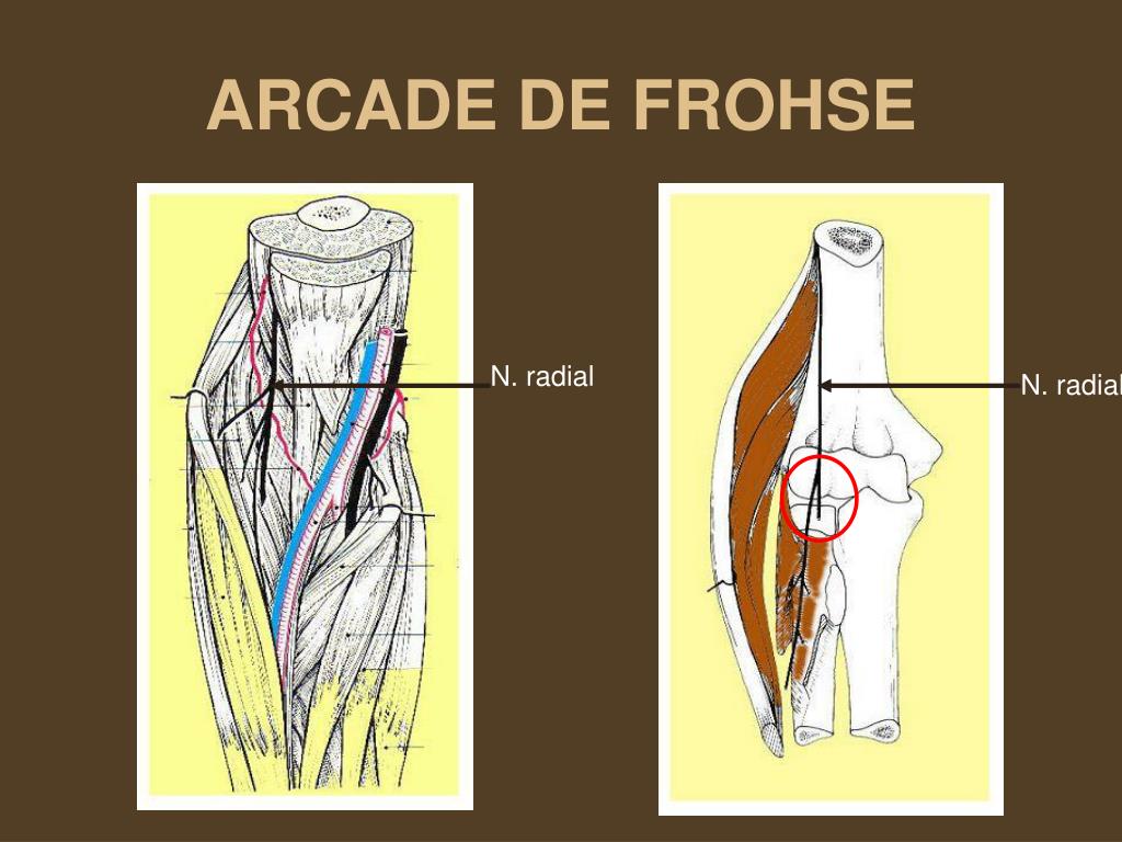
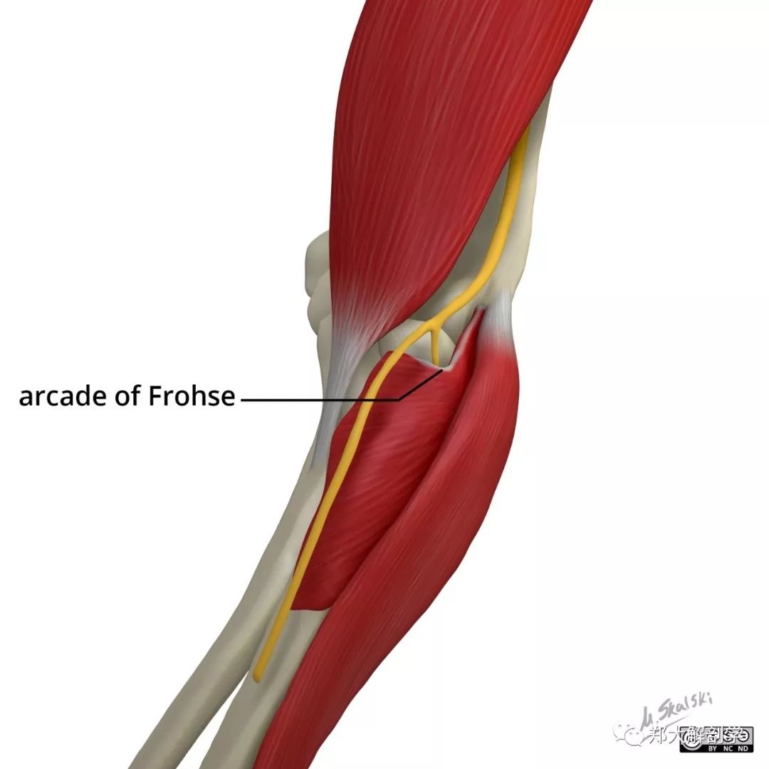
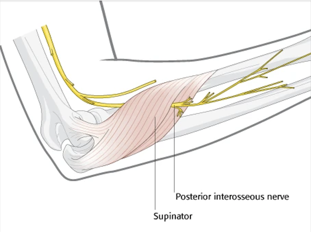
Citation, DOI, disclosures and article data. The arcade of Frohse (pronounced "\ˈfʁoːzə \") is also known as the supinator arch. The arcade is formed by a fibrous band between the two heads of the supinator muscle. The deep branch of the radial nerve passes beneath the arcade accompanied by vessels known as the leash of Henry.
PPT BELLAN Élodie DOLEAC Marie LEFRANCOIS Angélique ANNEE 2007 2008
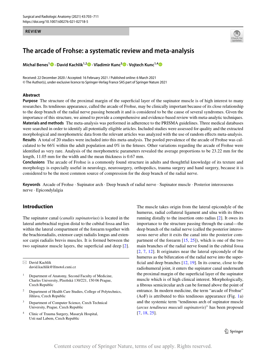
The arcade of Frohse is a commonly found structure in adults and thoughtful knowledge of its texture and morphology is especially useful in neurology, neurosurgery, orthopedics, trauma surgery and hand surgery, because it is considered to be the most common source of compression for the deep branch of the radial nerve. The structure of the proximal margin of the superficial layer of the.
胫神经,股,踝管(第2页)_大山谷图库
Pathology. It is a result of posterior interosseous nerve compression from trauma, micro-trauma, space-occupying lesions or inflammation. The most commonly described sites of compression are the arcade of Frohse and the distal edge of the supinator muscle respectively 2.Other potential sites (proximal to distal) are: fibrous bands anterior to the radiocapitellar joint

Figure 2 from Posterior Interosseous Nerve Palsy Caused by a Ganglion
The arcade of Frohse, the leash of Henry, and the entire supinator muscle should be divided to relieve PIN compression. The incision for the brachioradialis-splitting approach is slightly more anterior over the mobile wad. The brachioradialis fascia is divided, and muscle fibers split bluntly, gaining access to the supinator and PIN.

PIN Compression Syndrome Hand Orthobullets
The posterior interosseous nerve is much longer and enters the radial tunnel underneath a musculotendinous arch, the arcade of Frohse. The arcade of Frohse, which is the most common point of compression, is a connection between the deep and superficial heads of the supinator and is fibrotendinous in 30% to more than 80% of the population. [6]

Anatomy arcade of Frohse by posterolateral surgical approach
On physical exam, the patient has weakness of extension of the digits and wrist. Five potential sites of compression of the posterior interosseous nerve have been identified (7a,8a). Of these, the proximal tendinous edge of the supinator muscle (arcade of Frohse) is the most frequent site of posterior interosseous nerve entrapment (7a,8a) 2.
The arcade of Frohse a systematic review and metaanalysis Request PDF
We identified the Frohse arcade with a well-developed fibrous constitution in 22 of the 30 dissected limbs (73%) and of muscular constitution in 8 (27%). The distal margin of the supinator muscle.

Radial Tunnel Syndrome — ChiroUp
The arcade of Frohse is the most common site of compression and represents a thickened tendinous proximal edge of the superficial head of the supinator, whereas the normal edge is thin and membranous. The tendinous thickening is developmental, occurring in 30-100% of people, most likely due to repetitive pronation-supination .

IP Innovative Publication Pvt. Ltd
The PIN enters the radial tunnel underneath a musculo-tendinous arch, the arcade of Frohse. Formed by the upper free border of the superficial head of the supinator, the arcade of Frohse is a semicircular fibrous arch that remains fibrous medially and is found in 30-80% of anatomical specimens (Spinner, 1968, Clavert et al., 2009).

frohse’s arcade coachingultrasound
The arcade of Frohse is a commonly found structure in adults and thoughtful knowledge of its texture and morphology is especially useful in neurology, neurosurgery, orthopedics, trauma surgery and hand surgery, because it is considered to be the most common source of compression for the deep branch.
(PDF) A cadaveric study of the Arcade of Frohse
The arcade of Frohse. The AF is not a homogenous structure; its shape and the type of fibers are variable. Debouck and Rooze proposed a classification of the arcade based on its morphology . Type A indicates a resistant tendinous arcade. Type B is a mixed musculotendinous arch with two types of fiber alternating.

The drawing provides an anterior view of the course of the radial nerve
arcade of Frohse, distal edge of supinator. Radial nerve compression occurs in throwing and overhead activities. Repetitive pronation and supination can also cause similar symptoms. It is important to examine the cervical spine and the radial nerve both proximal and distal to the elbow. It has been postulated that a proximal compression can.

Figure 1 from Crosssectional sonographic assessment of the posterior
Radial Tunnel Syndrome is a compressive neuropathy of the posterior interosseous nerve (PIN) at the level of proximal forearm (radial tunnel). Diagnosis is made clinically with pain only (maximal tenderness 3-5 cm distal to lateral epicondyle) without any motor or sensory dysfunction. Treatment is a prolonged course of conservative management.

Ultrasound scanning for Arcade of Frohse YouTube
Case Discussion. Ultrasound demonstrated evidence of posterior interosseous nerve (PIN) entrapment at the arcade of Frohse (supinator muscle) caused by a band of scar tissue. In the setting of trauma to the wrist and hand, it was not possible to assess for finger drop and radial deviation of the wrist on extension.
Posterior Interosseous Nerve Injury to The Incarcerated forearm Dr
The arcade of Frohse is identified as a tendinous band at proximal side of supinator muscle. Initially the radial recurrent blood vessels (leash of Henry) should be ligated just proximal to the arcade of Frohse. The arcade of Frohse is released, and the superficial head of the supinator muscle is divided totally to ensure that the inferior.